Advantages of Fundus automated perimetry over Standard Automated Perimetry include the possibility to measure sensitivity at specific retinal locations, higher accuracy thanks to retinal-tracking based compensation of eye movements and the simultaneous assessment of function (expressed by retinal sensitivity) and structure (images of the ONH and of the retina).
Fundus automated perimetry provides a simultaneous, quantitative assessment of fixation characteristics.
COMPASS overcomes such limitations and brings visual field analysis to the next level!
In particular COMPASS, for the first time, extends field coverage to 30° + 30° and employs luminance parameters and a sensitivity scale as used in standard automated perimetry.
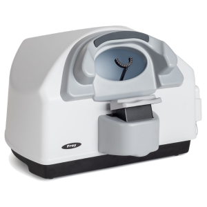
The first fundus automated perimeter capable of performing standard 24-2 visual fieldtesting and delivering true color confocal images.
Superior quality of color and red-free images
Retinal Tracking
Fixation Analysis In Glaucoma
Benefits
- High-resolution confocal imaging of the ONH and of the central retina
- Combined structure and function analysis
- Fully automated operation
- Comprehensive and clear printout
- Operator friendly
- More patient comfort: test can be suspendedat any time without data loss