Is specifically designed for vitreo-retinal specialists and ophthalmologists.
California includes a new UWF optomap® imaging modality; Indocyanine Green angiography (icg) while retaining:
- Composite color
- Red-free
- Autofluorescence (af)
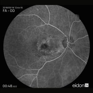
- Fluorescein angiography (fa)
BENEFITS
- California was designed as a compact, table-top model to reduce space requirements. further the new design leads to ease of use and faster image capture.
- Non-mydriatic high resolution imaging through many cataracts and/or 2mm pupils saves time in busy practices.
- Comprehensive retinal analysis through multiple wavelengths and image modalities, all in UWf.
- Composite color images can be viewed in their separate laser channels to show specific depths of the retina:
– Green (532 nm) “red-free” visualizes the
sensory retina to the RpE
– Red (635 nm) shows deeper structures
of the retina (RpE to choroid)
– infrared (802 nm) provides images at
the choroid level
– Blue (488 nm) is used during fa procedures - Innovative review platform that simplifies workflow while enabling comparison overlay between image modalities and over time.
- Browser based image review enables simple integration and easy access to your data from any connected pc or tablet in a hippa compliant environment.
- interweaved angiography enables parallel capture of fa and icg images without manually switching between imaging modalities.
- With UWf views of 200 degrees or up to 82% of the retina, eyecare professionals can see 50% more of the retina when compared to other conventional imaging devices1 .