AP-250 and AP-250BY use green color LED projection of stimulus in Goldman size III. AP-250BY additionally offers test Blue-on-yellow with a blue stimulus Goldman V size and yellow backlight in accordance with the requirements of the SWAP perimetry. The software supplied with the device offers a wide range of strategies, fields and test parameters. Control of fixation is performed automatically using the built-in camera or by controlling the position of the blind spot. Built-in data analysis include regression analysis and standardized formats for presenting and printing test results. Frey AP-250 as well as AP-250BY can be easily set up with any PC computer running the Windows operating system.
Static perimetry is the most frequently used technology for quality of visual field verification, detection of glaucoma and for monitoring glaucoma related changes in visual field. AP-250 and AP-250BY are supported by a rich library of test fields and strategies that meet and exceed user requirements and broad range of perimetry applications.
Bi-Driving is test for binocular patient examination with vision field extended to 80˚ nasally and temporally. The test fulfils requirements for driver testing. During the test, both eyes of the patient are simultaneously monitored by the digital camera.
Blue-on-Yellow static perimetry is a test used for early glaucoma detection. This feature is available on AP-250BY and performed according to SWAP recommended Goldman size V stimulus in blue color presented on yellow color background.
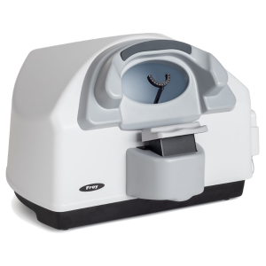
AP-250 and AP-250BY measurement bowl is very well designed and engineered housing a combination advanced digital bowl illumination controls which reduces requirements for test room illumination conditions. For improved patient comfort, ventilation is provided by whisper-quiet concealed fans.
Electrically driven chinrest with movement across horizontal and vertical axis, allows for easy patient positioning. Movement of the chinrest can be controlled with push buttons located on the front of the perimeter housing or from PC with perimeter software. Anatomical head and chin support provide patient comfort during the examination.
Network communication between PC and AP-250 and AP-250BY perimeters only require one USB 2.0 port, creating an effective and seamless interface with any PC running Windows. All PC network connected devices such as printers or storage servers can be used for examination test result printing or data backup and storage.
Regression analysis module allows user to track changes in field of vision in time, using easy for interpenetration graphs. Multiple examination results of the same patient can be used for the analysis. Regression curves can be presented i absolute dB values, in relation to hill of vision or age normative values or particular global parameters like PD or AD. Analysis of the data can be limited to particular area of vision field like periphery or center, or can cover entire tested field of vision.
To better visualize relation of vision field defects and retinal image, Fundus overlay function allows to import fundus image and then use it as a background for displaying values recorded during the examination. Proper position of fundus image in relation to perimetry data is achieved by selecting macula and blind spot on fundus picture.